Development and optimization of human gray matter microstructure quantification using diffusion MRI
Join me for a quick tour of my PhD research, whether you're a seasoned pro or just getting your feet wet
Modeling the Brain's Microstructures: Commonly Used Approaches in Diffusion MRI
In order to study the brain's microstructures using diffusion MRI, we need to use mathematical models to interpret the data. There are two main types of models that are commonly used in diffusion MRI: signal representation models and multi-compartment models.
Signal representation models, such as diffusion tensor imaging (DTI) and diffusion kurtosis imaging (DKI), do not make any assumptions about the underlying structure of the brain. Instead, they are based on the idea that the diffusion of water molecules can be described by a mathematical model that describes the movement of water molecules within the brain. These models can provide useful information about the brain's white matter tracts and the connections between different brain regions.
Multi-compartment models, such as the white matter tract integrity (WMTI-Watson), are based on the idea that the diffusion of water molecules is affected by different compartments within the brain, such as neurites, extra-cellular space, and cerebrospinal fluid (CSF). These models can provide more detailed information about the brain's microstructures and how they are connected, but they are also more complex and require more computational resources. They are often used to study specific types of tissue, such as white matter or gray matter, and can provide valuable insights into the structure and function of these tissues. By continuing to develop and optimize these models, we can gain a better understanding of the brain's microstructures and how they are connected, which could have important implications for the diagnosis and treatment of various brain disorders.
The Potential of a Grey Matter Model in Diffusion MRI
By developing and optimizing a grey matter model for Diffusion MRI, we could better understand the structure and function of grey matter, which could have important implications for the diagnosis and treatment of various brain disorders. For instance, research has shown that grey matter volume is reduced in some brain disorders, such as schizophrenia and Alzheimer's disease, and it may be that these changes could be detected in advance at the microscopic level. By studying grey matter, we could learn more about the underlying causes of these disorders and develop more targeted treatments. In addition, such a model could be used to study the effects of various interventions, such as medications or behavioral therapies, on grey matter. This could help us identify the most effective treatments for different brain disorders.
The Neurite Exchange Imaging model (NEXI)
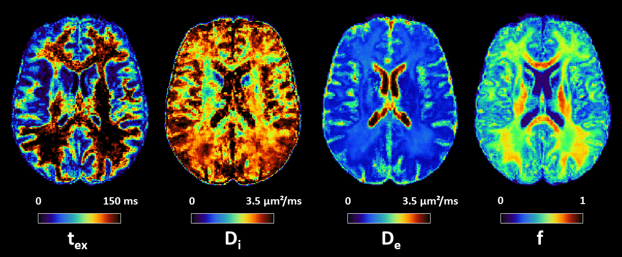
The Neurite Exchange Imaging (NEXI) model, proposed by my supervisor Dr. Ileana Jelescu and in parallel by Dr. Sune Jespersen and Jonas Olesen as SMEX (Standard Model with EXchange), was introduced recently to recognize and quantify water exchange across the neurite membrane. As such, NEXI is applicable on clinical grade scanners because it does not necessarily require short diffusion times. NEXI models the neurites as a collection of randomly-oriented sticks, occupying a relative signal fraction \(f\), where the intra-neurite diffusion is uniaxial with diffusivity \(D_{i,∥}\). Moreover, given the quasi-uniform orientation-distribution of neurites in gray matter, the extra-neurite compartment is considered to be Gaussian isotropic with characteristic diffusivity \(D_e\). The two compartments exchange with a characteristic time \(t_{ex}\). NEXI models the total orientation-averaged signal as the sum of these two exchanging compartments. They are assumed to have the same transverse relaxation time, or \(T_2\). The soma are not explicitly modeled and the signal contribution arising from this compartment is most likely pooled with the signal contribution from the extra-cellular space in NEXI.
My contributions so far
This section is still under development. If you want a more complete overlook on my research, please have a look at my posters from previous conferences.
NEXI estimation pipeline in python
My first contribution was to develop a rapid way of applying my model to both actual scanner data and synthetic data. I then published a simplified version on pypi to enable other researchers to use our model on their own data, whatever their acquisition sequence. The code is available here on GitHub, as well as here on pypi.
Quantifying human gray matter microstructure using NEXI and 300 mT/m gradients
This project focuses on the in-human implementation of NEXI on a 3T Connectom scanner and analysis of performance of different NEXI variants. The Connectom scanners are an important steppingstone in terms of hardware capabilities between preclinical MRI systems (with gradients >600 mT/m) and standard clinical MRI systems (with gradients ≤ 80 mT/m). They provide the opportunity of an initial translation of NEXI in human subjects by enabling the acquisition of the necessary broad range of b-values (0 - 7.5 ms/µm²) at diffusion times 20 - 49 ms, that are short enough to capture exchange processes with expected \(t_{ex}\)=10-50 ms.
Furthermore, the high data quality in terms of signal-to-noise ratio (SNR) is an opportunity to explore the performance of four NEXI fitting variants. I compared the two-compartment NEXI estimates to the ones from its three-compartment variant, allowing for an extra parameter capturing a possible 'dot' compartment, filled with stationary water. In the cerebellum, the presence of such a compartment has been shown (Tax et al., 2020), but its plausibility in the cortex had yet to be confirmed or refuted.
This research was the subject of a paper available on Imaging Neuroscience. More information will follow in the coming months.
Other subjects not yet published
Acquisition and processing of clinical MRI data (from the CHUV Prisma scanner)
Available soon. Preliminary version available in Biorxiv
Optimization of the protocol with Explainable AI and Self-Supervised Learning
Available soon. First apparition in the ISMRM 2025 Presentation : Reducing the NEXI acquisition time for the quantification of human gray matter microstructure on the CONNECTOM 2.0 scanner.
Extending gray matter models theory
Available soon. First apparition in the ISMRM 2024 Poster : the Generalized Exchange Model.